


Ear Examination
How to examine the ear: ENT exams for doctors, medical student finals, OSCEs and MRCP PACES
Introduction
- Wash hands
- Introduce self
- Explain examination and ask if the patient is in any pain
- Ask them to tell you if you cause them any discomfort with your examination
General Inspection
- If wearing a hearing aid, advise the patient to remove it
- Check for any asymmetry or if the patient presents with unilateral symptoms
- Any evident congenital facies (e.g. Down’s syndrome)
- Cauliflower ears may come from blunt trauma (sport contact)
- Look for specific abnormalities
- Tophi (gout)
- Sebaceous cysts
- Extra pinnae
- Skin tags or a pre-auricular sinus
- Scars
- Post auricular
- Endaural
- Discharge (e.g. wax or otorrhoea)
- Inflammation or ulceration
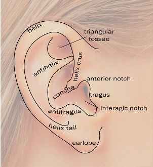
Surface anatomy of the ear
Palpate
- Pinnae
- Post auricular region
- Mastoid area
- Front of the tragus (ask patient to open and close mouth)
- Gently pull on the pinna (pain indicates there may be inflammation of the external auditory meatus [EAM])
Video on the ear examination and otoscopy
Otoscopy
- Hold the otoscope like a pen between the thumb and index finger
- Use your right hand for examination of the right ear and left hand for the left ear
- Slowly insert around 1-1.5cm just past the hair of the lateral canal
- Gently pull the pinna upwards, backwards and outwards
- Be careful! This may cause the patient discomfort if they have inflammation in their EAM
- Inspect the tympanic membrane
- If bulging it may lose its bony landmarks and usually is a sign of pus in the middle ear
- If retracted it will have accentuated bony landmarks and may signify a dysfunctional eustachian tube
- Inspect for:
- Discharge, scaling, inflammation, foreign bodies, stenoses, cerumen and exostoses
- Check drum to ensure:
- Not retracted
- No perforations
- Not bulging
- Colour and translucency

The tympanic membrane as seen through an otoscope
Extra tests
- Assess the patient’s hearing:
- Ask if they have any hearing loss
- Observe their ability to hear you during the examination
- If hearing loss is suspected then the tuning fork, Rinne and Weber’s tests may be useful
Conclude the exam
- Thank the patient
- Make sure they are comfortable
Click here to learn about the nose examination and here to learn about other ENT examinations
Perfect revision for medical student finals, OSCES and PACES