


Hand Examination
How to do a rheumatological hand examination – for doctors, medical student finals, OSCEs and MRCP PACES
Background
In the hand examination, as in any musculoskeletal examination think in terms of:
- Inspection (“look”)
- Palpation (“feel”)
- Manipulation (“move”) which comprises:
- Active movement
- Passive movement
- Stability
- Throughout these stages consider the hand in terms of its three basic components: skin, soft tissues and bones
Introduction (WIIPPPE)
- Wash your hands
- Introduce yourself (name and position)
- Identity of patient (confirm name and date of birth)
- Permission (consent and explain examination: “I’m going to examine your hands now, is that OK?”)
- Pain?
- Position
- Rest the patient’s arms on a pillow placed across their lap
- Expose
- You should be able to see both arms from fingers up to and including the elbow
- Ask them to remove any watches or bracelets
Inspection (“look”)
- Begin by examining the hands as they rest on the pillow on the patient’s lap
- Start with the dorsal aspect
- Ask patient to reveal the palmar surface and compare subtle differences that go undetected
- Ask the patient to show you their elbows
- For all three positions, systematically consider the components of the hand: skin, soft tissue and bone
Video on the hand examination
Skin
- Scars
- Scars over joints can indicate previous surgery on arthritic joints
- Inspect the anterior aspect of the hand for surgical scars from decompression of the carpal tunnel [click here for photo]
- Carpal tunnel syndrome may be secondary rheumatoid arthritis
- Skin quality
- Skin quality can be affected by both disease processes and treatments
- Systemic sclerosis (SS) causes tight and thickened skin tapered over the fingertips [click here for photo]
- Psoriasis (in psoriatic arthritis) give scaly silver plaques on extensor surfaces, particularly the elbows [click here for photo]
- Any vasculitis, including that secondary to RA or systemic lupus erythematosus (SLE) can cause finger-pulp infarcts
- Corticosteroids cause thinning of the skin and easy bruising
- Colour changes
- Redness may reflect underlying inflammation
- Raynaud’s phenomenon
- White (ischaemia); blue (cyanosis); red (reactive hyperaemia)
- It is important to establish the presence of Raynaud’s phenomenon if considering a diagnosis of SS, as its absence makes such a diagnosis highly unlikel (over 95% of patients with SS have Raynaud’s phenomenon)
- Longlasting blanching of the knuckles on clenching the fists is a pointer to Raynaud’s phenomenon even in a warm room
- Specific signs
- Gottron’s papules [click here for photo]
- Roughened red papules on the extensor surfaces of the fingers occur in dermatomyositis
- Telangiectasia [click here for photo]
- Especially if situated on palms and in nail folds are characteristic of systemic sclerosis
- Mechanics hands [click here for photo]
- This is a sign of thick, cracked skin over the tips and sides of the fingers and can be found in dermatomyositis and antisynthetase syndrome
- Gottron’s papules [click here for photo]
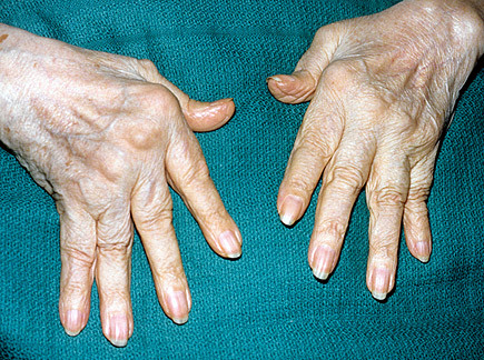
Classical hand changes in rheumatoid arthritis
Soft tissue
- Nails
- Psoriatic arthritis (PsA)
- 90% of patients with PsA will have typical nail changes including:
- Onycholysis (lifting from the nail bed) [click here for photo]
- Pitting [click here for photo]
- Longitudinal ridging [click here for photo]
- 90% of patients with PsA will have typical nail changes including:
- Systemic sclerosis and dermatomyositis
- Dilated nail fold capillaries [click here for photo]
- Other vasculitis
- Nails may reveal the nail fold infarcts of vasculitides [click here for photo]
- Psoriatic arthritis (PsA)
- Subcutaneous deposits
- Calcification in the pulp of the fingers is a feature of limited cutaneous systemic sclerosis
- Previously called CREST syndrome (Calcinosis, Raynaud’s phenomenon, Esophageal dysmotility, Sclerodactyly, Telangiectasia)
- White subcutaneous deposits located asymmetrically around joints are more likely to be tophi associated with gout [click here for photo]
- Note that tophi may occur at atypical sites (for example the fingertips)
- Calcification in the pulp of the fingers is a feature of limited cutaneous systemic sclerosis
- Soft tissue swelling
- Generalised puffiness is a common, non-specific sign in the early stages of connective tissue diseases.
- Synovial hypertrophy occurs in RA and noting the site of the inflammation is important for diagnosing complications. Diffuse cylindrical swelling of one or two digits (dactylitis), can be due to swelling specifically of the flexor tendon, as occurs in PsA (see Figure 5)
- Nodules
- May be apparent on inspection of the extensor surface of the elbows or other areas subject to external pressure, and suggest RA
- Muscle wasting
- May be caused by lower motor neuron lesions or reduced movement of damaged joints.
- Note the pattern of wasting e.g.:
- Nerve root [e.g. T1]
- Peripheral nerve [e.g. median]
- Muscle group [e.g. elbow flexors]
Common rheumatological causes of lower motor neuron lesions
| Mononeuropathies (involvement of one nerve) | Rheumatoid arthritis is commonest. Often affects median nerve (carpal tunnel syndrome) and ulnar nerve |
| Mononeuritis Multiplex (asymmetric peripheral nerve involvement) | Many, including: rheumatoid arthritis; diabetes; vasculitides (e.g. SLE; PAN; Wegeners) |
| Polyneuropathy (symmetric involvement of multiple nerves) | Amyloid; sarcoid |
Bone
- Many rheumatological diseases show a predilection for certain joints whist sparing others
- Inspect systematically and to compare both hands
- Each inflamed joint should be assessed for current inflammation or inactive processes, which can be achieved through palpation
 Usual joint distribution of osteoarthritis (OA) and rheumatoid arthritis (RA)
Palpation (“feel”)
- Again, move through skin, soft tissue and bone
Skin
- Temperature
- Localised warmth can indicate inflammation, whereas cold extremities may be caused by Raynaud’s phenomenon.
- Increased temperature is best felt by palpating across the area with the back of the hand. A hot, painful erythematous joint must be considered septic until proven otherwise and accordingly managed as an emergency. Remember that septic joints are not always single (up to 20% have multi-joint involvement)
- Sensation
- Test in terms of distal neuropathy and individual nerve function
- Distal neuropathy
- Begin by testing for loss of sensation in the fingertips (peripheral neuropathy; a feature of RA)
- Individual peripheral nerve
- The sensory distribution of the median, ulnar and radial nerves should be evaluated as RA may also produce mononeuropathies through compression or, more rarely, vasculitic involvement of the vasa nervorum
- Median: palmar surface of the index finger
- Ulnar: over the little finger
- Radial nerve: base of the thumb
- The sensory distribution of the median, ulnar and radial nerves should be evaluated as RA may also produce mononeuropathies through compression or, more rarely, vasculitic involvement of the vasa nervorum
Soft tissue
- Pitting oedema
- Bogginess of subcutaneous tissues overlying a joint is a sign of acute inflammation and may occur in early rheumatoid arthiritis
- Bursae or effusions
- These may produce a tense swelling around the joint. These can be distinguished from other enlargements by the ability to squeeze fluid from one side of the joint to the other
- Tendons
- Can become palpably thickened and nodular in early-stage RA, sometimes preceding the deforming joint changes
- Joint capsule thickening
- This is confined to the anatomical boundaries of the joint. It feels doughy and can be best felt by pinching the soft tissues gently and rolling the synovium over the joint
Bone
- Gently squeeze across the joints in a methodical order, assessing for the nature of any swelling present
- Remember that inflamed joints are painful and must be examined gently.
- As with the abdominal examination, look at patient’s face if possible while palpating to discern any pain
- Each metacarpophalangeal (MCP) and interphalangeal (IP) joint must be palpated in turn and compared to the opposite side. This is best done by holding the joint between your thumb and forefinger and palpating gently
- If the swollen joint feels soft, warm and diffusely tender it is usually due to acute synovitis. If it is hard it is usually due to bony overgrowth

Psoriatic arthritis with dactylitis
Manipulation (“move”)
- By this stage it is likely you will have a good idea of the diagnosis. Manipulation is useful to confirm your suspicions but particularly to get an idea of how the patient’s condition impacts on them functionally
Active movement
- Ask about any pain the patient feels, particularly with which movements
- Pain in most or all directions is the most sensitive sign of synovitis; pain in one plane of movement is more characteristic of a localised intra- or peri-articular lesion.
- Ask about stiffness
- This is related to fluid retention in the peri-articular tissues and is an indication of the inflammatory processes
- Function
- The key to testing active movement is to work out what the patient can and can’t do functionally. Ask what their pain or stiffness stops them doing. To test this more fully you can them ask them to:
- Squeeze your finger (fully flex the hand powerfully)
- Pick up a coin from a table (pincer grip)
- Pretend to lock a door with a key (key grip)
- Do up a button (coordination and fine motor control)
- These movements are common in every day life so are a useful gauge of what the patient can and can’t do
- The key to testing active movement is to work out what the patient can and can’t do functionally. Ask what their pain or stiffness stops them doing. To test this more fully you can them ask them to:
Passive movement
- Watch the patient’s face while moving their joints so as not to take the range of movement beyond the active range
- During the movement crepitus may be palpated (the sensation of creaking) and can be a sign of damage to the bearing surfaces. Characteristically crepitations are coarse in OA and fine in RA
Stability
- Joint stability is provided through a combination of dynamic stability (muscle power) and static stability (ligaments and intact joint surfaces). The stability of the MCP joints should be assessed if RA is suspected, as these frequently sublux in advanced RA
- Dynamic stability is measured by provideing resistance at the proximal phalanx of each finger in turn, whilst the patient attempts to extend the MCP joint
- In advanced rheumatological diseases weakness and atrophy may be apparent
- Static stability is measured through gently stressing the joint in directions controlled by a ligament, feeling for any laxity of sudden step as the joint subluxes
Closure
- Thank patient, ensure they’re comfortable and ask if they need any help in getting dressed
- Wash hands
- Turn to examiner, hands behind back, holding stethoscope, before saying: “To complete my examination, I would like to…”
- Further examinations:
- Other joints
- Respiratory (if concern about rheumatoid arthritis, which is associated with pulmonary fibrosis and pleural effusions)
- Abdomen (if concerned about rheumatoid arthritis [splenomegaly in Felty’s sundrome])
- Further investigations:
- Bloods
- FBC (anaemia of chronic disease)
- U&Es (renal impairment with vasculitis and for drug dosing)
- LFT (baseline, pre-medication)
- ESR and CRP (correlates with disease activity in rheumatological conditions)
- TFT (if autoimmune concern)
- Urate (if considering gout)
- Immunology
- Rheumatoid factor
- Does not rule RA in or out
- Positive in up to 20% of normal population
- Also positive in other autoimmune disease e.g. Sjogrens, SLE, autoimmune liver disease
- Negative in around 30% of patients with RA, may become positive later on in disease course
- ACPA (anti cyclic citrullinated peptide)
- Does not rule RA in or out but more specific than RF and predictor of poor prognosis
- HLA-B27
- Rheumatoid factor
- Hand XR
- To look for characteristic erosions
- Joint aspiration
- If effusion and concern about diagnosis or septic process
- Bloods
- Further examinations:
Click here to learn about the skin examination
…and click here to learn about the diagnosis and management of rheumatoid arthritis
Perfect revision for medical student finals, OSCES and PACES