


Lumbar puncture (LP)
Anatomy in lumbar puncture (LP)
- CSF
- Produced in choroid plexus in both lateral ventricles
- 20mls/ hour or 500mls/ day
- Found in subarachnoid space
- Total volume <1/3 that of daily production
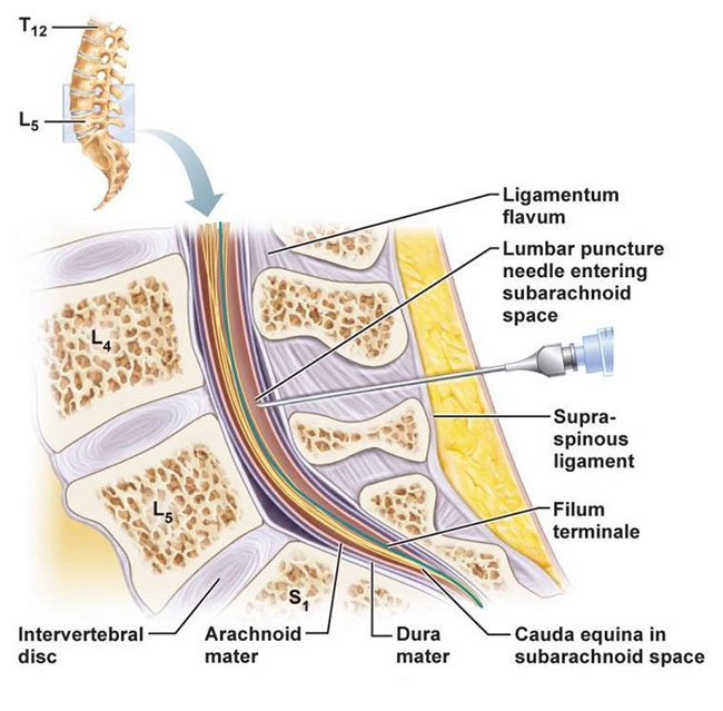
- Spinal anatomy
- Spinal cord ends at distal end of L1
- LP preferentially performed at L4,5 or L3,4
- Posterior Superior Iliac Crest (PSIS) located at L4,5
- Place palms of hands over PSIS so that superior edge is under your index finger. Your thumbs will connect at, or point to, the approximate location of the L4 vertebrae
- Approximate LP depth(cm) = 1 + 17 x weight (kg) / height (cm)
- Layers penetrated by spinal needle
- Skin; supraspinous ligaments; interspinous ligaments; ligamentum flavum; epidural space; dura; subarachnoid membrane into subarachnoid space
Indications for lumbar puncture (LP)
- Suspected meningitis
- Suspected subarachnoid haemorrhage
- Fever of unknown origin
- CNS leukaemia or lymphoma
- Evaluation of neurological conditions eg. recurrent seizures/ multiple sclerosis
- Diagnosis and treatment (therapeutic pressure reduction) in raised intracranial pressure
Contraindications to lumbar puncture (LP)
- Presence of infection at lumbar puncture site
- Papilloedema or signs raised intracranial pressure
- Severe thrombocytopaenia
- Uncorrected bleeding disorders
- Presence of cerebral mass lesions
- e.g. Abscesses, tumours, intracranial haemorrhage, subdural haematomas
Possible complications of lumbar puncture (LP)
- Post-LP headache
- Infection
- Spinal cord injury and bleeding into spinal canal (very rare)
- Failure to find subarachnoid space
Pre-procedure
- Assess risk of raised intracranial pressure
- Any evidence on imaging?
- Any false localising signs noted (e.g. unexplained third or sixth nerve palsy)?
- Check platelets and clotting reasonable
- Ensure absence of infection or metalwork at lumbar puncture site
- Check for allergies to latex, betadine, lignocaine or other medications
- If doing the LP for possible subarachnoid haemorrhage, ensure 12 hours have passed since the onset of symptoms
- Document informed consent (written if possible)
- Infection, bleeding, failure, pain
- Damage to surrounding structures (including nerves and vessels)
- Damage to cord incredibly rare. Warn patient they may feel a shooting pain down one of their legs as the needle goes in and to tell you if they do so.
- Headache (in up to one third of patients)
- Usually resolves within a couple of hours with simple analgesia. Advice on hydration, caffeine intake and lying flat for one hour post-procedure (though no good evidence for this)
Equipment required for lumbar puncture (LP)
- Dressing trolley & sharps bin
- Sterile field
- Sterile dressing pack and gloves
- 2% Chlorhexadine swabs
- Analgesia
- 4mls of 1% or 2% Lidocaine
- Orange (25G) needle (x1)
- Green (19G) needle (x1)
- 5ml Syringe (x1)
- Gauze swabs and small dressing
- Spinal needle (atraumatic needles reduce headache)
- Manometer
- Familiarise yourself with this first and ensure it turns freely
- Four universal CSF sample containers (labelled 1-4). Usually yellow-topped.
- One glucose tube (fluoride oxalate tube, often grey)
Positioning for lumbar puncture (LP)
- A lumbar puncture can be performed in two positions:
- Lateral recumbent
- Patient on his or her side with head propped up on a single pillow to keep spine straight
- Knees & torso flexed to optimise interlaminar foramen of vertebrae
- Ask or use assistant to draw patient’s legs up to their chest
- Ensure craniospinal & transverse planes remain stable
- NB. excessive flexion can compromise upper airway
- Sitting
- Useful in patients with pulmonary disorders or potential airway compromise
- Seat patient on edge of bed
- Flex trunk by having patient lean forward & rest elbows on table or on knees
- NB. CSF pressure cannot be reliably measured in this position
Procedure
- Create your sterile area and put on sterile gloves
- Identify & sterilise needle insertion site
- Mark entry point with blunt end of needle
- Draw up lignocaine in 5ml syringe
- With an orange (25G) needle raise intradermal wheal
- With a green (22G) needle advance into subcutaneous tissues
- Always check for entrance to blood vessel prior to injection of lignocaine & never inject into spinal canal
- Wait a few minutes for lignocaine to work. During this time do a final check of your equipment and make sure you have an assistant to help with bottles
- Insert spinal needle
- Make sure stylet in place before advancing needle
- Advance needle slowly in direction of umbilicus with bevel facing upwards (towards the ceiling) if patient in lateral position
- Advance slowly through spinous ligament resistance until you feel some give (sometimes described as a “pop”) with change in resistance as needle enters subarachnoid space
- Remove stylet and check for flow of spinal fluid.
- If no flow, replace stylet, rotate spinal needle a few millimetres & then recheck
- If obstructed, or if needle meets resistance, withdraw the needle with stylet in place, recheck position & re-attempt the procedure
- With flow of CSF, attach manometer & 3-way tap to the needle
- Turn the 3-way tap upwards to allow CSF fluid to fill the manometer. Once the CSF has stopped advancing (leaving a swinging menincus) remember this CSF pressure.
- A normal opening pressure is 12-20 cmCSF
- Once pressure read, collect samples by placing sterile containers under the 3-way tap and turning it to let the CSF directly out
- Make sure bottles are in the correctly numbered sequence
- Usually 1-2ml each container
- If getting cytology up to 20ml is required (the more CSF, the higher the sensitivity)
- Turn the 3-way tap upwards to allow CSF fluid to fill the manometer. Once the CSF has stopped advancing (leaving a swinging menincus) remember this CSF pressure.
- Once all samples are taken withdraw needle
- There is no need to replace the stylet to do this
- Note that in cases of doing a lumbar puncture for raised ICP it may be necessary to take a closing pressure as well as an opening pressure
- Place bandage over needle insertion site
- Ensure samples correctly labelled, phone laboratory staff inform them of urgent sample to be processed
- If necessary take them to the laboratory yourself
- Samples usually sent for:
- Cell count
- Gram stain, culture & sensitivity
- Protein, glucose
- Oligoclonal bands (OCB) if indicated
- Virology panel (PCR for HSV, VZV at the very least)
- Cytology (consider spare sample to keep in medical fridge)
- Spectrophotometry if looking for subarachnoid haemorrhage
- The ‘three tube test’ (a decrease in red cells in consecutive tubes on visual inspection) is notoriously unreliable
- Take blood samples for glucose and oligoclonal bands (if indicated) immediately post-LP
- For how to interpret CSF studies, see: ‘How To Interpret CSF studies post-LP’
NEJM video on how to perform a lumbar puncture
Post-procedure
- Check for headache and ensure PRN simple analgesia prescribed
- Advise on good hydration and taking a caffeine-containing drink to decrease headache
- Conventionally the patient should lie flat for at least one hour post-procedure though there is no good evidence that this reduces headache
- Remember to take blood samples for glucose +/- oligoclonal bands immediately post-LP
Top Tips for lumbar punctures (LPs)
- Take time to explain the procedure well. Patients have heard of LPs and so are often scared of them – a reassuring explanation goes a long way.
- Ask the patient to tell you if they feel a shooting pain down one leg as the needle is going in. If they do, readjust your angle of entry to aim more towards the other side.
- Ensure you locate the correct space in the midline. In very obese patients this may be difficult. Asking the patient whether or not you are pressing in their midline can be helpful. They will know even if you don’t!
- Before the procedure make sure you are happy with how the manometer and 3-way tap connect. This is the most complex part of the procedure and needs to go as smoothly as possible. Try the tap prior to the procedure – it often sticks so can be difficult to release first time round.
- If you are unsure what samples need to be sent, always take a spare tube and ask the lab to save the sample.
Click here for medical student OSCE and PACES guide to interpreting CSF
Common CSF analysis examples for medical students, finals, OSCEs and MRCP PACES